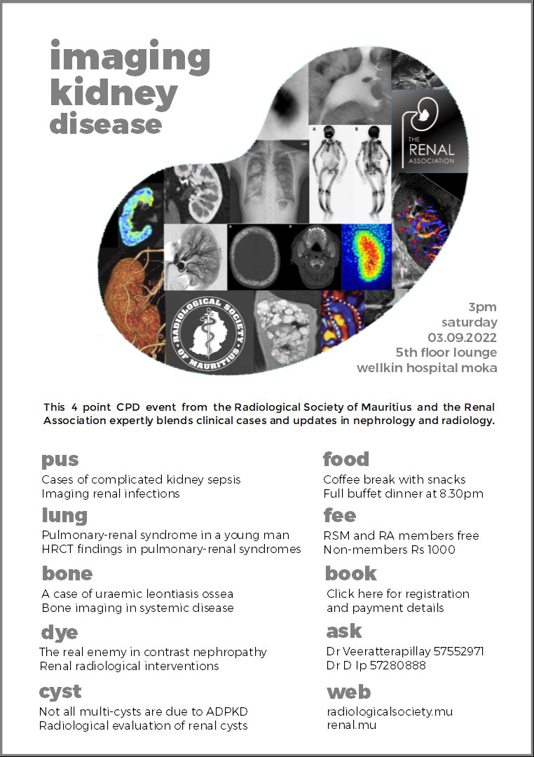
IMAGING KIDNEY DISEASE SYMPOSIUM 2022
SATURDAY 3 SEPTEMBER 2022
5th FLOOR, WELLKIN HOSPITAL - REDUIT, 3PM
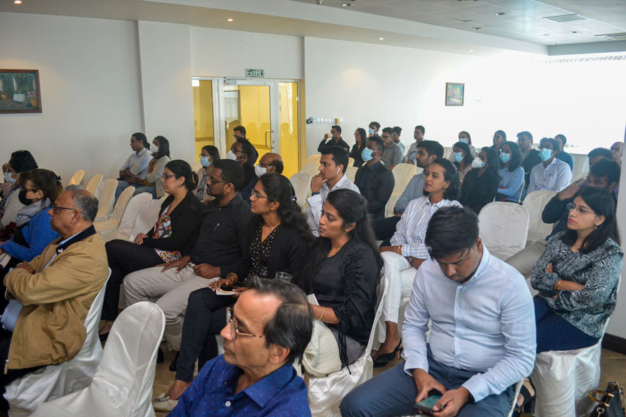
This joint Radiological Society of Mauritius and Renal Association symposium was held on 3 September 2022. This is the first collaboration between the two societies and it was both well attended and received.
Scientific Programme
1Cases of complicated kidney sepsis - Dr A.S. FEDALLY
This a presentation of recent cases of kidney sepsis in local hospitals highlighting the presentation, the diagnostic challenges, the protracted clinical course, complications and the severe attendant morbidity and mortality. There will be a review of the epidemiology and the evidence base for clinical management in the scientific literature. The high prevalence of diabetes in Mauritius and the ageing population suggest that kidney sepsis will become ever more prevalent and a case will be made for a better national strategy to combat this scourge.
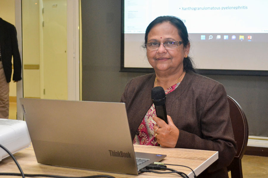
2Imaging renal infections - Dr A. VEERATTERAPILLAY
Infections of urinary tract is a common problem. Imaging is required in certain cases with poor response to treatment and find causes which needs alternative management. Talk will discuss different types of pyelonephritis like emphysematous and xanthogranulomatous pyelonephritis and the diverse imaging findings in these cases. Imaging is essential in diagnosis of complications of renal infection like renal and perirenal abscess which are often seen due to large number of diabetic patients in Mauritius.
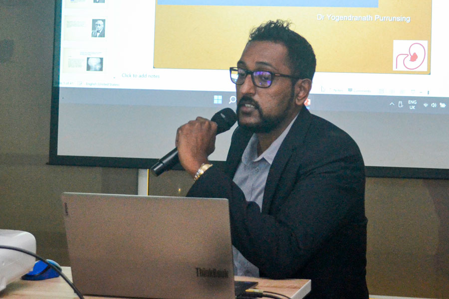
3A case of uraemic leontiasis ossea - Dr Y. PURRUNSING
Secondary hyperparathyroidism (SHPT) is a long‐term complication of chronic kidney disease–mineral and bone disorder. SHPT is characterized by hyperplasia of the parathyroid glands and abnormal secretion of parathyroid hormones (PTH), calcium and phosphorous metabolic disorders, renal osteodystrophy, vascular and soft tissue calcification, malnutrition, and other multiple system complications. SHPT seriously affects the quality of life, increases cardiovascular disease risk and mortality rate. Uraemic leontiasis ossea is a rare manifestation of SHPT causing craniofacial bone deformity accompanied by lesions of the nerve, cardiovascular, respiratory, bone, or other systems within the body. The case discussed in this presentation involves a 62‐year‐old male haemodialysis patient suffering from leontiasis ossea, pectus excavatum, vascular calcification, spontaneous bone fractures, and lower limb deformities.
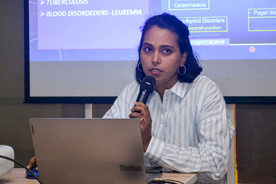
4Bone imaging in systemic disease - Dr D. GHOORAH JEVALLEE
Bone is a dynamic organ of the endoskeleton, maintaining structural integrity, mineral reservoirs and blood production.For patients affected by these processes, radiologic imaging plays a central role in diagnosis, monitoring treatment, and risk stratification. Radiologists need to be aware of the imaging findings to wisely guide the best practice of medicine.This lecture focusses on several key types of metabolic bone disease: osteoporosis, osteomalacia, scurvy, renal osteodystrophy, hyperparathyroidism, Paget’s disease etc These diseases all affect bone however with shared characteristic features that can be discerned through imaging.
5Renal radiological interventions - Dr T. GHOORAH
This session will discuss the various tools and techniques utilized by the radiologists to reach the diagnosis and also the key role played by various newer interventional techniques in the treatment of the various pathologies in urology and nephrology patients. Local practices will be compared to those abroad as well the effectiveness and safety of the newer radiological interventional techniques over the more established surgical techniques.
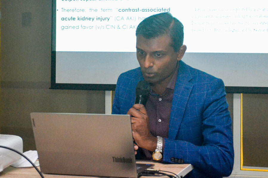
6The real enemy in contrast nephropathy - Dr O. BHEEKHARRY
Contrast-associated acute kidney injury (AKI) features a decrease in kidney function within days after the intravascular administration of iodinated contrast material. In the 1950s, a high incidence of AKI was initially reported in patients with pre-existing kidney disease who undergoing intravenous pyelography with contrast agents. Over time, development of new contrast agents, recognition of risk factors, and implementation of preventive care have reduced AKI rates to the point that recent studies now suggests that the AKI risk is exaggerated. Does it now mean that potentially life-changing angiographic procedures may be denied to patients with chronic kidney disease because of concern about precipitating AKI? This presentation summarizes the pathophysiology of contrast-associated AKI, the diagnostic criteria, and risk stratification; discusses current controversies regarding the incidence of this condition and preventive care.
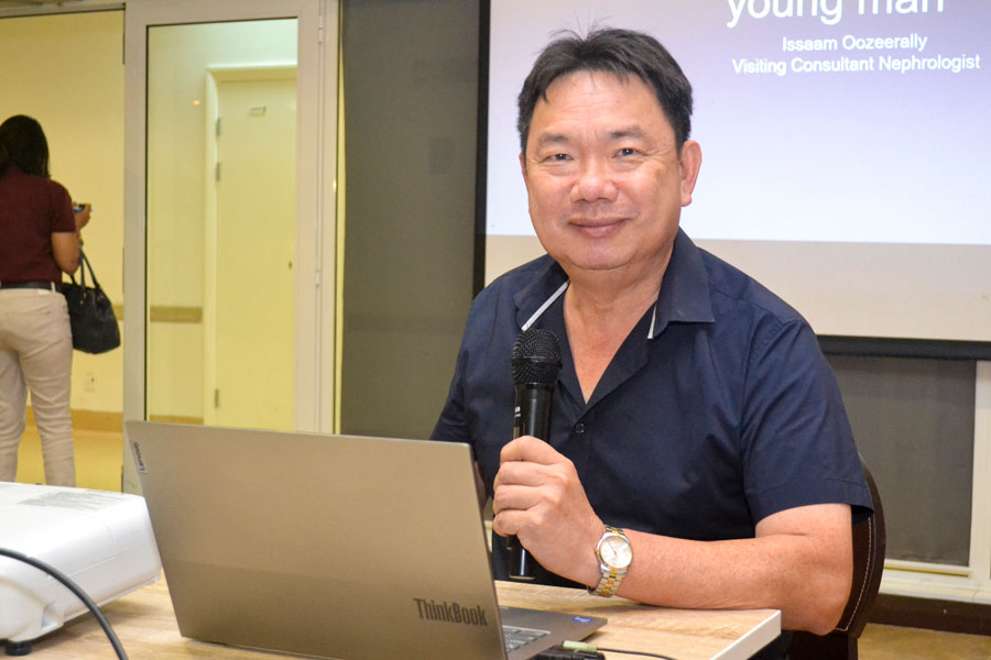
7Pulmonary-renal syndrome in a young man - Dr I. OOZEERALLY
The talk will be a case presentation of a 40 year old male who was diagnosed with a pulmonary-renal syndrome. The latter is a rare diagnosis which, as the name suggests, is a disease process affecting both lungs and both kidneys. Complications include pulmonary haemorrhage requiring invasive ventilation and rapidly progressive glomerulonephritis causing acute kidney injury and needing emergency haemodialysis. Mortality is very high especially with pulmonary involvement. A review of the epidemiology, clinical features, diagnostic tests and evidence-based treatment will follow the case presentation.
8HRCT lungs in pulmonary-renal syndromes - Dr P. CHUI WAN CHEONG
Pulmonary Renal Syndrome is rare with a high rate of morbidity and mortality. There is respiratory failure due to diffuse alveolar haemorrhage and renal failure due to glomerulonephritis. The lecture aims to demonstrate high resolution CT findings of the lungs in diffuse alveolar haemorrhage. From an acute episode to chronic changes in the lungs after repeated alveolar haemorrhage.
9Radiological evaluation of renal cysts - Dr R. GOLAMAULLY
Cystic renal lesions are commonly encountered on radiologic examinations. Complex and multifocal cystic renal lesions are often a diagnostic challenge, since they can represent neoplastic and non-neoplastic conditions. Not all multi-cysts are due to autosomal dominant polycystic kidney disease (ADPKD), a hereditary form of adult cystic renal disease and by far the most common hereditary cause of end stage renal failure (ESRF). The Bosniak classification system is a well-established imaging method, which helps radiologists and surgeons in daily practice in the differentiation of nonsurgical from surgical lesions.
10Not all multi-cysts are due to ADPKD - Dr D. IP
This session will explore the diagnostic challenges in the evaluation of patients with multiple cysts through a series of clinical case presentation. Autosomal dominant polycystic kidney disease (ADPKD) is the commonest genetic disorder worldwide and is, unsurprisingly, often given as a diagnostic label to any patient with multiple cysts. The clinical features and management of ADPKD will be reviewed. There are many other polycystic diseases all linked by numerous extra-renal manifestations and being various forms of ciliopathy.
Download Event Poster
Imaging Kidney Disease Poster